CBS 2019


CBSMD教育中心


首页


CBS 2019
关于CBS


会议通知
会议组织
主办单位
南京市心血管病医院
亚洲分叉病变俱乐部
南京医科大学附属南京医院
南京市第一医院医学发展医疗救助基金会
协办单位
中华医学会心血管病学分会
国家卫生健康委能力建设和继续教育心血管病学专家委员会(CBCEC)
美国心血管造影和介入学会
亚洲心脏学会
北京合众关爱心脏健康基金会
联系我们
境内代表注册管理:
彭红娟 : +86-13913895477
徐海梅 :+86-13914736846
 cbsnj@cbsmd.cn
境外代表注册管理:
林玲 : +86 (25) 52271398
+86-13951884596
cbs@cbsmd.cn
病例征集管理:
赵莹莹 :+86-13815408517
sub@cbsmd.cn
地址:
 江苏省南京市秦淮区长乐路68号,南京市心血管病医院9号楼905区心内科办公室
 210006


主席团
主席团
大会主席
联合主席


教育中心
教育中心
• 线上教育 ( 注册后可使用个人定义界面搜集科研图书馆文献、下载教育资源、参与在线病例讨论。)
• 教育资源


• 科研动态


科研图书馆
科研图书馆

科研文章
> 急性冠脉综合征
> 动脉粥样硬化性心血管疾病
> 分叉支架
> 心脏肿瘤科
> 充血性心力衰竭
> 双重抗血小板治疗持续时间
> 药物涂层球囊
> 血流储备分数
> 血管内超声指导
> 光学相关断层扫描
> 肺动脉高压
> 旋磨术
> 血流动力学与动脉粥样硬化
> 左主干支架
> 经导管主动脉瓣置换
> 经皮左心耳封堵
> 二尖瓣/三尖瓣瓣膜
> 推荐文献
荐读文献
Management of Acute Myocardial Infarction During the COVID-19 Pandemic
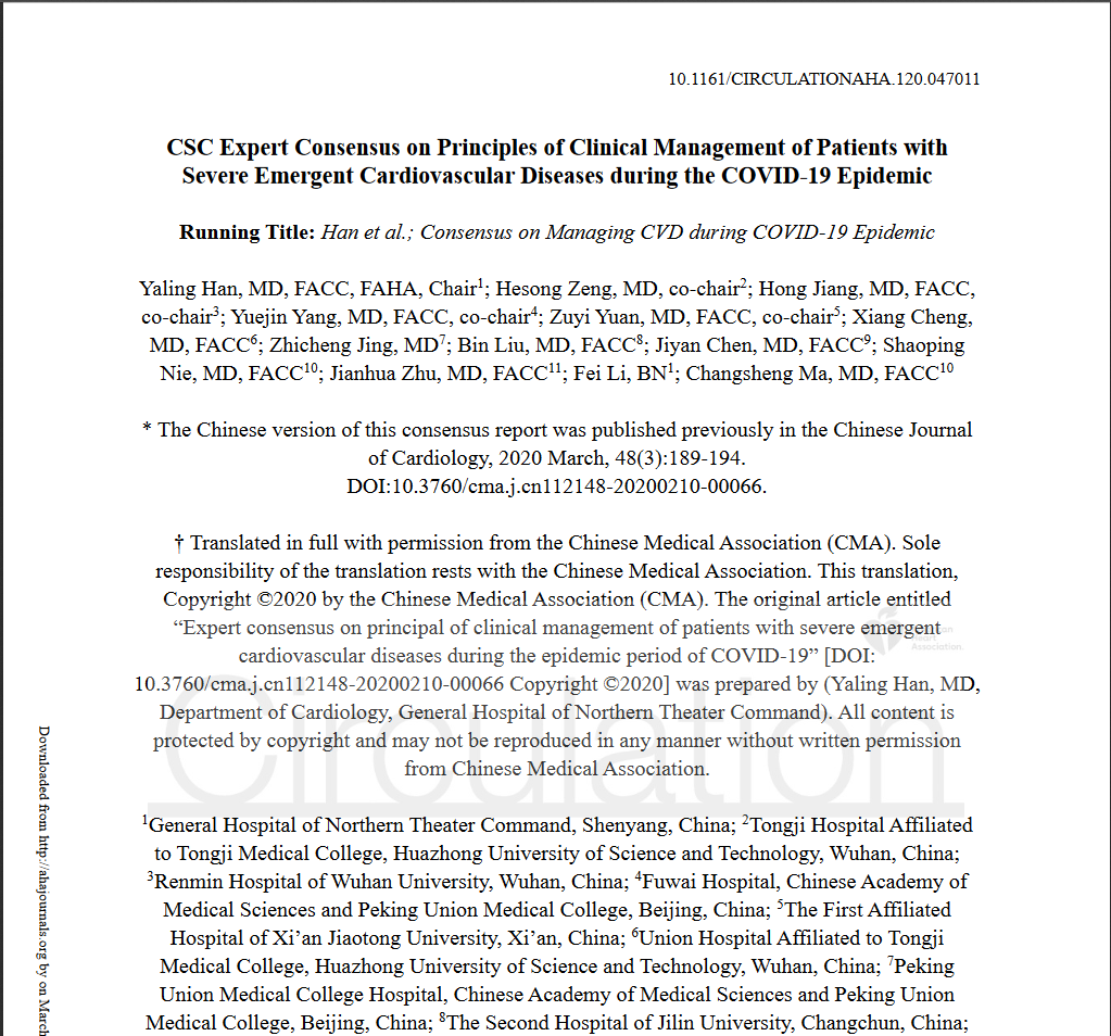
CSC Expert Consensus on Principles of Clinical Management of Patients with Severe Emergent Cardiovascular Diseases during the COVID-19 Epidemic
ACC临床简报:新型冠状病毒对心脏的影响(2019-nCoV)
SCAI Expert Consensus Statement Update on Best Practices for Transradial Angiography and Intervention
急性冠脉综合征
Original Research2021 Apr, 77 (13) 1599–1611
JOURNAL:J Am Coll Cardiol. Article Link
S Torii , Y Sato , F Otsuka et al. Keywords: calcified nodule; acute coronary thrombosis; ACS
BACKGROUND - Calcified nodule (CN) has a unique plaque morphology, in which an area of nodular calcification causes disruption of the fibrous cap with overlying luminal thrombus. CN is reported to be the least frequent cause of acute coronary thrombosis, and the pathogenesis of CN has not been well studied.
OBJECTIVES - The purpose of this study is to provide a comprehensive morphologic assessment of the CN in addition to providing an evolutionary perspective as to how CN causes acute coronary thrombosis in patients with acute coronary syndromes.
METHODS - A total of 26 consecutive CN lesions from 25 subjects from our autopsy registry were evaluated. Detailed morphometric analysis was performed to understand the plaque characteristics of CN and nodular calcification.
RESULTS - The mean age was 70 years, with a high prevalence of diabetes and chronic kidney disease. CNs were equally distributed between men and women, with 61.5% of CNs found in the right coronary artery (n = 16), mainly within its mid-portion (56%). All CNs demonstrated surface nonocclusive luminal thrombus, consisting of multiple nodular fragments of calcification, protruding and disrupting the overlying fibrous cap, with evidence of endothelial cell loss. The degree of circumferential sheet calcification was significantly less in the culprit section (89° [interquartile range: 54° to 177°]) than in the adjacent proximal (206° [interquartile range: 157° to 269°], p = 0.0034) and distal (240° [interquartile range: 178° to 333°], p = 0.0004) sections. Polarized picrosirius red staining showed the presence of necrotic core calcium at culprit sites of CNs, whereas collagen calcium was more prevalent at the proximal and distal regions of CNs.
CONCLUSIONS - Our study suggests that fibrous cap disruption in CN with overlying thrombosis is initiated through the fragmentation of necrotic core calcifications, which is flanked—proximally and distally—by hard, collagen-rich calcification in coronary arteries, which are susceptible to mechanical stress.


 English









 CBS 2019
 CBS 2019 主席团
 教育中心
教育中心 科研动态
科研动态


