CBS 2019


CBSMD教育中心


首页


CBS 2019
关于CBS


会议通知
会议组织
主办单位
南京市心血管病医院
亚洲分叉病变俱乐部
南京医科大学附属南京医院
南京市第一医院医学发展医疗救助基金会
协办单位
中华医学会心血管病学分会
国家卫生健康委能力建设和继续教育心血管病学专家委员会(CBCEC)
美国心血管造影和介入学会
亚洲心脏学会
北京合众关爱心脏健康基金会
联系我们
境内代表注册管理:
彭红娟 : +86-13913895477
徐海梅 :+86-13914736846
 cbsnj@cbsmd.cn
境外代表注册管理:
林玲 : +86 (25) 52271398
+86-13951884596
cbs@cbsmd.cn
病例征集管理:
赵莹莹 :+86-13815408517
sub@cbsmd.cn
地址:
 江苏省南京市秦淮区长乐路68号,南京市心血管病医院9号楼905区心内科办公室
 210006


主席团
主席团
大会主席
联合主席


教育中心
教育中心
• 线上教育 ( 注册后可使用个人定义界面搜集科研图书馆文献、下载教育资源、参与在线病例讨论。)
• 教育资源


• 科研动态


科研图书馆
科研图书馆

科研文章
> 急性冠脉综合征
> 动脉粥样硬化性心血管疾病
> 分叉支架
> 心脏肿瘤科
> 充血性心力衰竭
> 双重抗血小板治疗持续时间
> 药物涂层球囊
> 血流储备分数
> 血管内超声指导
> 光学相关断层扫描
> 肺动脉高压
> 旋磨术
> 血流动力学与动脉粥样硬化
> 左主干支架
> 经导管主动脉瓣置换
> 经皮左心耳封堵
> 二尖瓣/三尖瓣瓣膜
> 推荐文献
荐读文献
Management of Acute Myocardial Infarction During the COVID-19 Pandemic
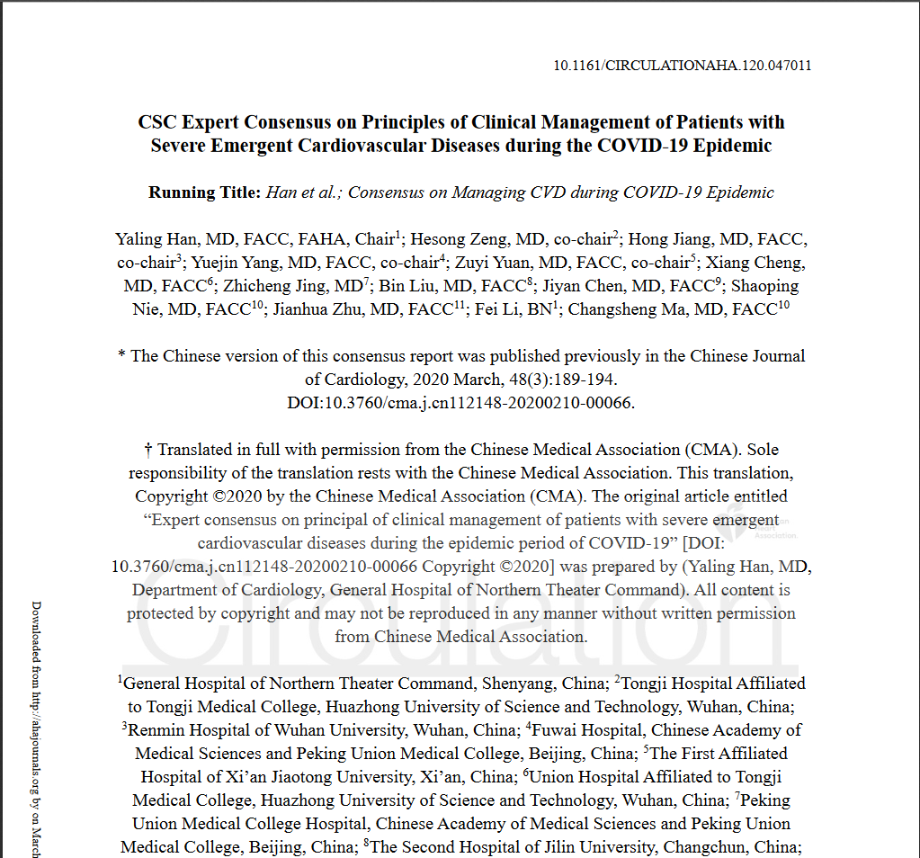
CSC Expert Consensus on Principles of Clinical Management of Patients with Severe Emergent Cardiovascular Diseases during the COVID-19 Epidemic
ACC临床简报:新型冠状病毒对心脏的影响(2019-nCoV)
SCAI Expert Consensus Statement Update on Best Practices for Transradial Angiography and Intervention
科学研究
Original Research2022 Feb, 79 (4) 341–351
JOURNAL:J Am Coll Cardiol. Article Link
G Masini , FJ Graham, P Pellicori et al. Keywords: chronic HF; iron deficiency; prevalence; prognosis


 English




 CBS 2019
 CBS 2019 主席团
 教育中心
教育中心 科研动态
科研动态


