CBS 2019


CBSMD教育中心


首页


CBS 2019
关于CBS


会议通知
会议组织
主办单位
南京市心血管病医院
亚洲分叉病变俱乐部
南京医科大学附属南京医院
南京市第一医院医学发展医疗救助基金会
协办单位
中华医学会心血管病学分会
国家卫生健康委能力建设和继续教育心血管病学专家委员会(CBCEC)
美国心血管造影和介入学会
亚洲心脏学会
北京合众关爱心脏健康基金会
联系我们
境内代表注册管理:
彭红娟 : +86-13913895477
徐海梅 :+86-13914736846
 cbsnj@cbsmd.cn
境外代表注册管理:
林玲 : +86 (25) 52271398
+86-13951884596
cbs@cbsmd.cn
病例征集管理:
赵莹莹 :+86-13815408517
sub@cbsmd.cn
地址:
 江苏省南京市秦淮区长乐路68号,南京市心血管病医院9号楼905区心内科办公室
 210006


主席团
主席团
大会主席
联合主席


教育中心
教育中心
• 线上教育 ( 注册后可使用个人定义界面搜集科研图书馆文献、下载教育资源、参与在线病例讨论。)
• 教育资源


• 科研动态


科研图书馆
科研图书馆

科研文章
> 急性冠脉综合征
> 动脉粥样硬化性心血管疾病
> 分叉支架
> 心脏肿瘤科
> 充血性心力衰竭
> 双重抗血小板治疗持续时间
> 药物涂层球囊
> 血流储备分数
> 血管内超声指导
> 光学相关断层扫描
> 肺动脉高压
> 旋磨术
> 血流动力学与动脉粥样硬化
> 左主干支架
> 经导管主动脉瓣置换
> 经皮左心耳封堵
> 二尖瓣/三尖瓣瓣膜
> 推荐文献
荐读文献
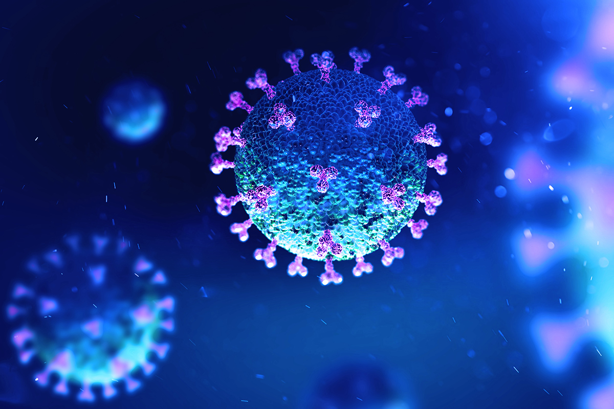
Management of Acute Myocardial Infarction During the COVID-19 Pandemic
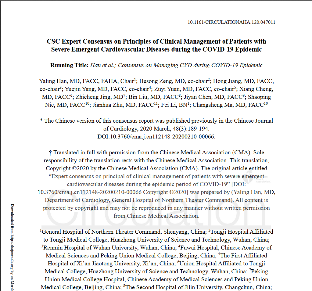
CSC Expert Consensus on Principles of Clinical Management of Patients with Severe Emergent Cardiovascular Diseases during the COVID-19 Epidemic
ACC临床简报:新型冠状病毒对心脏的影响(2019-nCoV)
SCAI Expert Consensus Statement Update on Best Practices for Transradial Angiography and Intervention
科学研究
Original Research2019 Mar 8. [Epub ahead of print]
JOURNAL:JACC Cardiovasc Imaging. Article Link
Jackson R, Al-Hussaini A, Adlam D et al. Keywords: intracoronary imaging; optical coherence tomography; spontaneous coronary artery dissection
OBJECTIVES - This study used optical coherence tomography to investigate the mechanism of false lumen (FL) formation in spontaneous coronary artery dissection (SCAD) by studying: 1) differences between fenestrated and nonfenestrated SCAD; 2) vasa vasorum density; and 3) light attenuation characteristics of the FL.
BACKGROUND - SCAD is an increasingly recognized cause of acute coronary syndromes, characterized by FL formation and compression of the true lumen (TL). The mechanisms underlying FL formation remain poorly understood.
METHODS - A total of 65 SCAD patients (68 vessels) who underwent acute OCT imaging as part of routine clinical care were included. Images were classified by the absence or presence of a connection (fenestration) between the TL and FL. Indexed measurements of TL stenosis, external elastic lamina (EEL) area, FL area, and light attenuation of the FL were assessed. Vasa vasorum densities of SCAD cases were compared with those in control non-SCAD myocardial infarction cases.
RESULTS - In nonfenestrated cases, there was significantly larger expansion of the EEL area (9.1% vs. -1.9%; p <0.05) and a larger FL area (73.6% vs. 53.2%, respectively; p <0.05) in dissected segments. No significant differences were found between vasa vasorum density in SCAD and those in control subjects. The FL contents were heterogeneous but attenuated less light than whole blood or thrombus (4.28 ± 0.55 mm-1 vs. 5.08 ± 0.56 mm-1; p < 0.05; vs. 4.96 ± 0.56 mm-1; p < 0.05).
CONCLUSIONS - These observational data suggest that the absence of a fenestration leads to increased FL pressure and compression of the TL. Although vasa vasorum may still be implicated in pathogenesis, increased vasa vasorum density could be an epiphenomenon of vascular healing.
Copyright © 2019 American College of Cardiology Foundation. All rights reserved.


 English




 CBS 2019
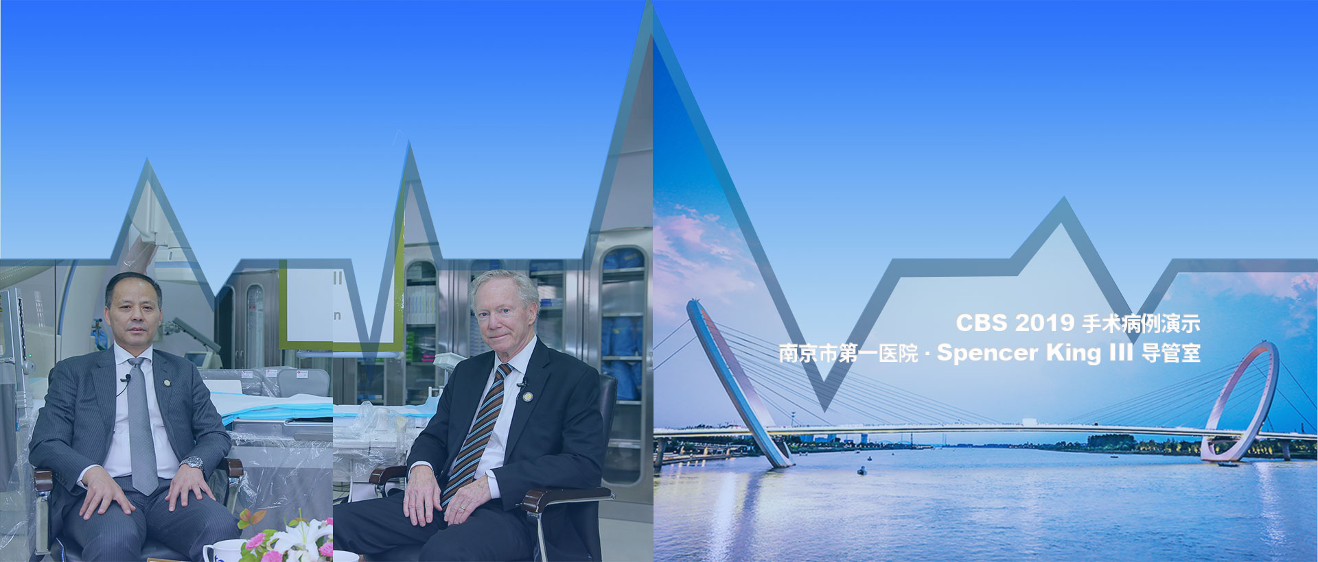
 CBS 2019 主席团
 教育中心
教育中心 科研动态
科研动态


